What are different MRI types
There are three types of magnetic resonance imaging methods, and although they serve different purposes, all three are used to visualize various aspects of the body’s anatomy:
- Magnetic Resonance Imaging (MRI) is often used to locate general abnormalities in the body. MRI shows detailed images of the body’s organs and tissues. MRI is a non-invasive medical imaging technique that shows precise details of body parts, especially of soft tissues. The MRI scanner uses strong magnetic fields, magnetic field gradients, and radio waves to generate images of the organs in the body. MRI is often used for disease detection, diagnosis, and treatment monitoring.
- Magnetic Resonance Angiography (MRA) is a specific type of MRI exam that allows doctors to look specifically at the arteries, without seeing all the overlying tissues. MRA sometimes might require the injection of a contrast material.
- Magnetic Resonance Venography (MRV) is an exam that uses an injection of a contrast material to improve the visibility of the veins without obstruction from overlayered tissues.

What to expect during an MRI
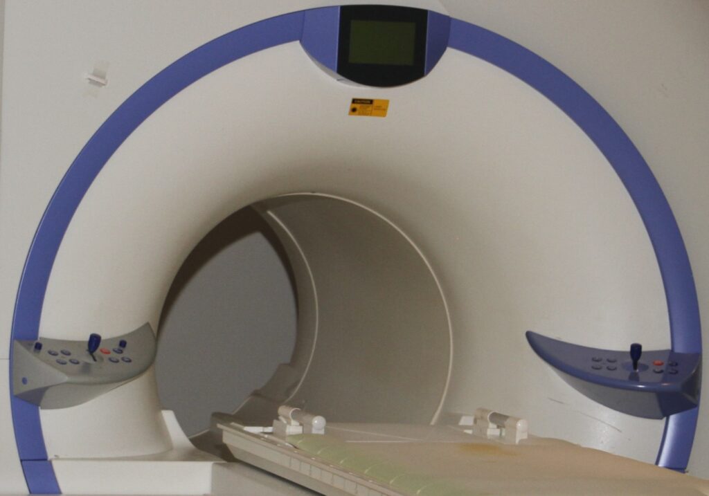
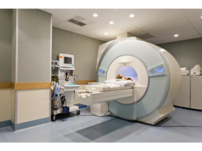
Both ends of an MRI machine are open. After you lay on a moveable bed, it will go in and out of the MRI machine. Remain steady and do not move as the bed moved back and fort under the MRI operation.
The MRI machine is a bit noisy, so the technician will provide you with a headset and a microphone so you can communicate with each other during the procedure. Again, it is critical that you do not move at all while the machine is taking images of your body part(s).
MRI is procedure are not painful, especially if the contrast injection is not required. Do tell the technician if you feel any discomfort of feel claustrophobic before the procedure begins.
MRI vs CT & PET
Lastly, an MRI does not involve X-rays or the use of ionizing radiation. Such methods are called computed tomography (CT) and positron emission tomography (PET) scans and are vastly different than an MRI.
Precautionary Screening
Precautionary screening is a crucial aspect of an MRI procedure to keep a patient safe and unharmed. Doctors refer patients for MRI procedures because they feel scan results give pertinent information about their patients. If you have any questions about your procedure, do not hesitate to ask your doctor and our technicians. You may not qualify for an MRI if you have any of these following conditions:
- Pacemaker
- Stent
- Pregnant
- Recorder
- Aneurysm Clip
- Pain Pump
- Shrapnel/ Bullets
- Pacing Leads
- Metallic Implants
- Defibrillator
- Neurostimulator
MRI Body Parts – with/ without contrasts
Pituitary, Head-Brain, Posterior Fossa, Sinuses, Orbits, IACs, Neck (soft tissue), Chest, Brachia Plexus, Cervical-Spine, Thoracic-Spine, Lumbar-Spine, Liver, Abdomen, Sacrum, SI Joint, Pelvis, MR Urogram, MRCP, Toe, Heel, Ankle, Foot, Tibia/Fibula, Femur, Knee, Hip, Hand, Wrist, Elbow, Humerus, Forearm, Clavicle, Shoulder, Scapula, and TMJ.
General Radiography
Skull Series, Nasal Bones, Facial Bones, Sinuses, Orbits, Mandible, Chest, Scoliosis Series, Skeletal Survey, Cervical-Spine, Thoracic-Spine, Lumbar-Spine, Obstructive Series, Sacrum, Pelvis, KUB, Abdomen, Bone Age, Toe, Heel, Ankle, Foot, Tibia/Fibula, Femur, Knee, Hip, Ribs, Finger, Hand, Wrist, Elbow, Humerus, Forearm, Clavicle, Shoulder, Scapula, and TMJ.
Report Result Availability
The exam results are typically available after radiologist interpretation, possibly 24 to 48 hours following your exam. Please be advised that due to HIPAA compliance regulations, we will not permitted to read the results over the phone. Of course, the results are also generally available to your referring doctor about 1 to 2 business days after most exams.


 Premier MRI Patient Instructions